MECHANISM OF INJURY
Fracture of both medial and lateral malleoli can be produced either by inversion or by eversion force. When the ankle-joint is exposed to strain, only one malleolus is fractured, but the continuance of the violence produces a fracture of the other malleolus.
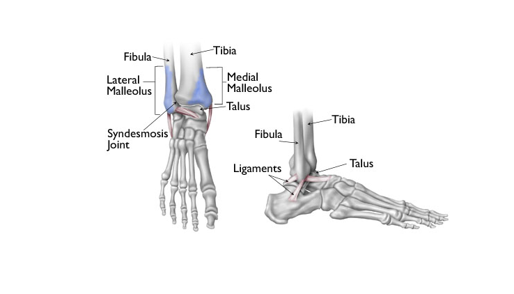
NATURE OF FRACTURE
The line of fracture of the fibula may be high up. When the stress is severe the interosseous tibiofibular ligament may be ruptured. The talus is shifted medially in adduction injury, whereas there is a lateral shift of talus by abduction or eversion violence.
DIAGNOSIS
Clinical features give an idea about the nature of the injury.
X-ray: This confirms the amount of talar shift and the presence of nay diastasis of the inferior tibiofibular joint.
TREATMENT
Bimalleolar fracture is unstable and surgery is often necessary.
- Closed reduction: Closed reduction is done with the patient lying on the table with the legs hanging over the edge. Displacement of the talus and malleoli are corrected by the application of pressure. A short leg plaster is applied, and reduction is checked by x-ray.
- Open reduction: Internal fixation is performed by inserting bone screw through separate incisions over medial and lateral sides of the ankle-joint. Bone screws can be accessible from the top orthopaedic implants suppliers company in India.
TRIMALLEOLAR FRACTURE
This is the extreme lesion of the ankle-joint produced by the external rotational injury. The third malleolus produced by the posterior-inferior part of the tibia is fractured.
NATURE OF FRACTURE
Several external rotation forces enable the talus to be rotated laterally and displaced posteriorly.
Stages of lesions produced: The fractures are produced in stages as the tension gradually rises with the increase of external rotation force.
- Lateral malleolus: There is a fracture of the lateral malleolus because of external rotation. This happens at the initial stage of violence.
- Medial malleolus: Medial ligament is put to severe stretch and fracture of the medial malleolus is produced. In some cases, it is only the medial ligament which is ruptured, and the medial malleolus does not suffer any fracture.
- Trimalleolar fracture: Lower and posterior margin of the distal end of the tibia is also known as the third malleolus. As the talus moves posteriorly, the extreme range of lateral rotation produces a vertical fracture of the posterior margin of the tibia. The fragment involves the articular surface of the tibia which is dis- placed posteriorly.
TREATMENT
Conservative treatment: Many [cases can be reduced by the closed method. Anatomical alignment is essential. Failure to achieve. this requires open. reduction. The procedure is done under general anesthesia. The patient lies on the table with the legs extended.
- Correction of posterior displacement: The heel is grabbed by one hand and pressed anteriorly while the distal end of the leg is pressed posteriorly by the other hand.
- Correction of lateral displacement: Pres- sure is now applied on the lateral malleolus directing medially while the distal end of the leg is pushed laterally.
Immobilization: Short leg plaster is applied, and the leg is kept elevated on a pillow while in bed. The patient can walk with crutches when the swelling disappears. No weight bearing is allowed for a period of 4-6 weeks. The plaster is removed after 10 weeks’ time.
Operative Reduction: Internal fixation is required when the anatomical reduction is not achieved. Internal fixation needs some Orthopedic Implants and surgical instruments used by the orthopedic surgeons. Plaster immobilization is done and managed in the same way as the closed reduction.


